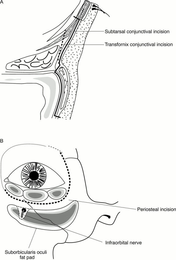
Figure 4 .

(A) Diagram showing transconjunctival approach to the inferior orbital rim and suborbicularis oculi fat (SOOF). The dissection continues posterior to the SOOF in the subperiosteal plane. (B) Diagram of left orbit showing location of SOOF in relation to the infraorbital nerve. The SOOF usually lies just above the infraorbital rim, but is shown lower here, mimicking involutional or paralytic changes.
