
Figure 2 .
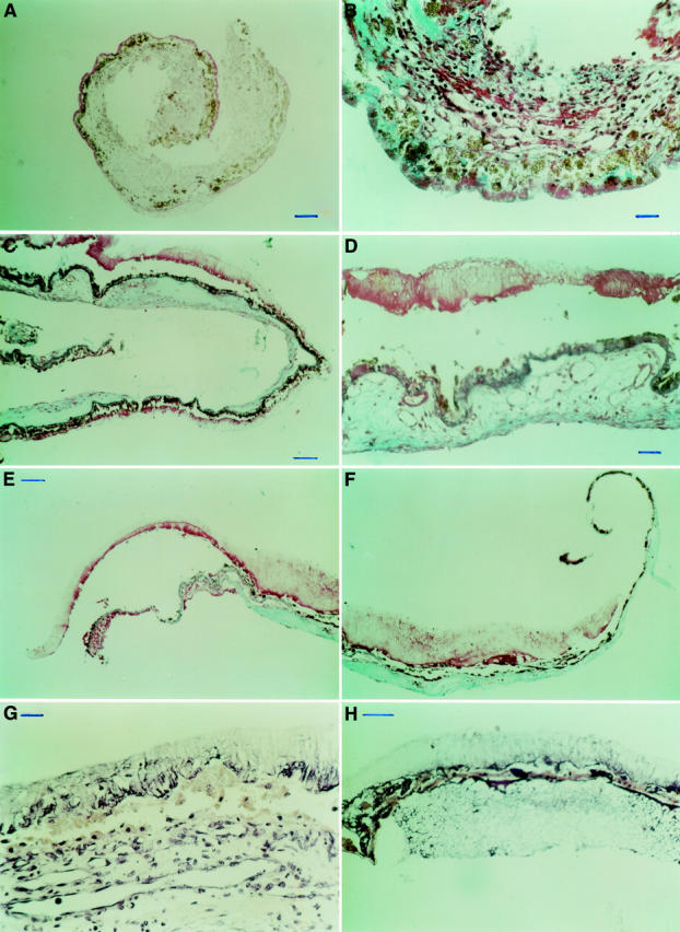
(A) PAS stain of a classic membrane, corresponding to Figure 1A and B, indicating a major subretinal and a minor sub-RPE fibrovascular component. Both these components are separated by diffuse drusen that stained as a PAS positive line. Bar = 100 µm. (B) MTC stain, detail of (A). Bar = 25 µm. (C) MTC stain of an occult choroidal neovascular membrane, corresponding to Figure 1C and D, shows a sub-RPE fibrovascular membrane and remains of outer segments covering the inner surface of the membrane. Bar = 100 µm. (D) MTC stain, detail of (C). Bar = 25 µm. (E, F) MTC stain of another occult choroidal neovascular membrane; fibrovascular tissue is seen at the choroidal side of the retinal pigment epithelium and remains of outer segment material cover the inner surface of the membrane. Bar = 100 µm. (G) PTAH stain of an occult choroidal neovascular membrane. Fibrin is seen interspersed with remains of outer segments. Bar = 25 µm. (H) PTAH stain of an occult choroidal neovascular membrane. A serofibrinous pigment epithelial detachment is seen. Bar = 50 µm.