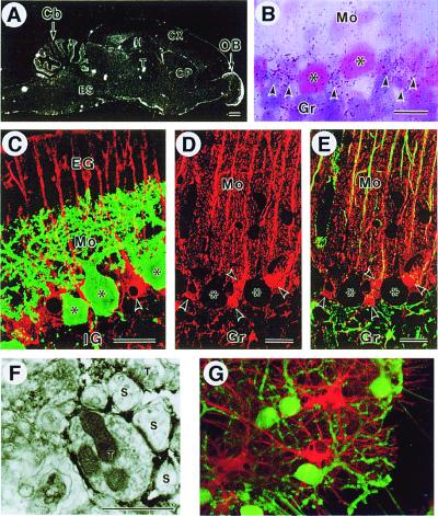
Figure 4.
3PGDH expression in the cerebellar cortex. (A) 3PGDH mRNA expression in the adult rat brain. Note intense expression in the cortical regions of the cerebellum (Cb) and olfactory bulb (OB). No signals are seen with a sense probe (data not shown). BS, brainstem; CP, caudate-putamen; Cx, cerebral cortex; H, hippocampus; T, thalamus. (B) Emulsion autoradiography for 3PGDH mRNA in the cerebellar cortex. Silver grains (arrowheads) were clustered around somata of PN (asterisks). Gr, granular layer; Mo, molecular layer. (C) Double immunofluorescence for 3PGDH (red) and calbindin (green) in the cerebellar cortex on the postnatal day 10. Note 3PGDH localization in the Bergmann glia (arrowhead), but not in PN (asterisks). EG, external granular layer; IG, internal granular layer. (D and E) Confocal images for 3PGDH (red) without (C) or with (D) simultaneous detection of GFAP (green) in the cerebellar cortex. Arrowheads indicate cell bodies of the Bergmann glia. Note colocalization of both immunoreactivities in Bergmann fibers running in the molecular layer, yielding an orange-to-yellow fusion color. (Scale bars: A, 1 mm; B–E, 20 μm) (F) Immunoelectron microscopy for 3PGDH in the molecular layer. Dark immunoreaction products are seen in lamellate processes of the Bergmann glia, which seal the synaptic cleft between spines of PN (S) and nerve terminals (T). (Scale bar indicates 1 μm.) (G) 3PGDH expression in cultured astroglial cells. Cerebellar mixed cultures were stained for 3PGDH (red) and calbindin D-28K (green) on DIV10. 3PGDH-positive cells were GFAP-positive astroglial cells (data not shown).

