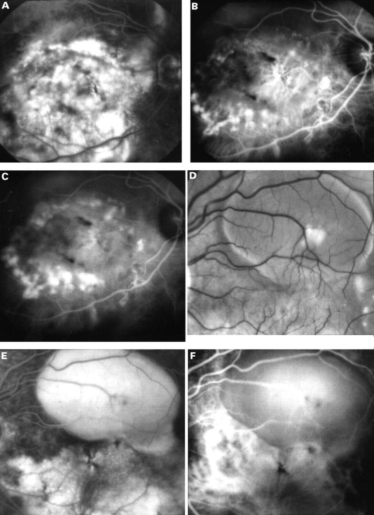
Figure 4 .

(A) A late fluorescein angiogram of the right eye of a 54 year old white man with counting fingers vision showing diffuse areas of hyperfluorescence between the temporal vascular arcades corresponding to a large serous elevation of the retina with subretinal fibrosis, hyperpigmentary changes, and subretinal and intraretinal haemorrhages. (B) Early ICG angiogram showing a ring of polyp-like aneurysmal dilatations of the choroidal vasculature in the macular region. In addition, there appears to be a choroidal neovascular complex centrally. (C) The mid-phase ICG showing staining of these polypoidal lesions. (D) In his left eye there was a pigment epithelial detachment. (E) Fluorescein angiography showed filling of the detachment and diffuse hyperfluorescence below and nasal to the lesion. (F) ICG angiography revealed a large diameter vessel complex at the same site with terminal dilatation of the vessels.
