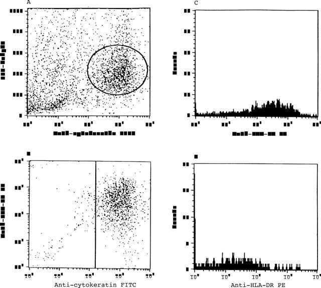
Figure 1 .
Cells isolated from imprint of ocular surface were incubated with a combination of CD3, anticytokeratin antibodies, and anti-HLA-DR antibodies. Cells were gated on a forward scatter cell size (FCS) versus side scatter (SSC) plot. CD3 positive cells and cytokeratin positive cells were separately analysed for the anti-HLA-DR molecule expression. (A) Epithelial cells were gated on a FSC versus SSC plot. A gate was set in to obtain HLA-DR positive epithelial cells. (B) Double gated cells were analysed for the HLA-DR molecule expression. (C) Epithelial cells with higher mean fluorescence intensities. (D) Epithelial cells with weaker mean fluorescence intensities.

