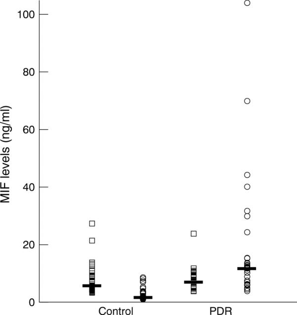
Figure 1 .
Macrophage migration inhibitory factor (MIF) levels in the vitreous and paired serum samples from eyes with macular hole or idiopathic epiretinal membrane (controls: n=41) and proliferative diabetic retinopathy (PDR: n=32). Open circles represent vitreous levels and open squares represent serum levels. The horizontal lines indicate the median concentration in each group. The vitreous MIF levels in PDR were significantly greater than levels in the controls (p<0.0001). Vitreous MIF levels were significantly higher than serum MIF levels in PDR (p=0.0026), but significantly lower than serum levels in the controls (p<0.0001).

