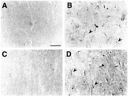
Figure 1.
High-magnification photomicrographs showing IL-1β (A and B) and IL-1Ra (C and D) immunoreactivity in the hippocampus of representative B6/CBA F1 mice locally injected with 0.08 nmol bicuculline methiodide (B and D) compared with vehicle-injected controls (A and C). A–D depict corresponding areas of the molecular layer of the dentate gyrus in the injected hippocampus. IL-1β (B) and IL-1Ra (D) staining was enhanced in cells with glial morphology 2 and 4 h after bicuculline injection respectively (arrowheads). No staining was apparent in vehicle-injected mice (A and C) or in mice receiving heat-inactivated cytokines (not shown). A similar immunocytochemical pattern of induction was observed after bicuculline-induced seizures in wild-type SV/129 littermate mice of IL-1R-type I knockout mice (see Table 2) compared with their respective vehicle-injected controls. (Bar = 100 μm.)

