
Figure 3 .
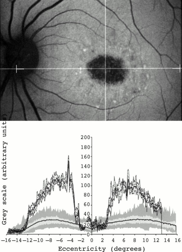
Fundus autofluorescent image of a 35 year old woman with Stargardt macular dystrophy-fundus flavimaculatus (top). An area of decreased signal at the centre of the macula and small foci of increased signal within it and around it were seen. Fundus autofluorescence profile showed very high background fundus autofluorescence levels across the entire area studied. Values of fundus autofluorescence at the fovea were within the lower normal values and peaks of increased autofluorescence at that site were also detected (bottom).