
Figure 4 .
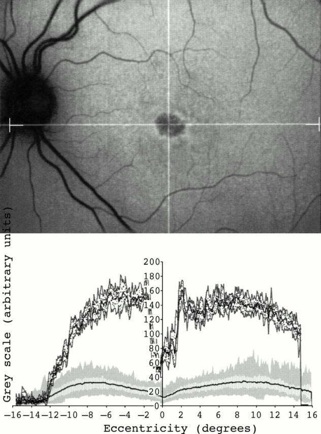
Fundus autofluorescent image of a 28 year old female with a bull's eye macular dystrophy (top). No focal areas with high or low intensity signal were detected outside the fovea in the cSLO image. Fundus autofluorescence profile demonstrated very high levels of fundus autofluorescence throughout the posterior pole (bottom).