Abstract
AIM—To assess uniformity of the corneal endothelial cell mosaic in children. METHODS—36 healthy children (5-11 years old, 16 boys, 20 girls) were assessed by specular microscopy. Endothelial cell density (ECD) was calculated from measured cell areas, and the number of sides/cell noted. RESULTS—Average values for ECD and cell areas were 3987 cells/mm2 (95% CI 3806 to 4168 cells/mm2) and 278 (SD 85) µm2 respectively, with normal distribution (COV 28.2%, range 17.4 to 39.2%) and with the average percentage of six sided cells being 66.6% (8.8%). Cell area was positively correlated to number of cell sides (p <0.01, r2=0.993), but the percentage of six sided cells was negatively correlated to ECD (p <0.01, r=0.493). CONCLUSION—A high ECD occurs in children, but this does not mean there is a high percentage of "hexagons".
Full Text
The Full Text of this article is available as a PDF (155.5 KB).
Figure 1 .
Typical non-contact specular micrograph of the corneal endothelium of a child. The ECD is 4050 cells/mm2 compared with the group averaged value of 3987 cells/mm2 (see Results).
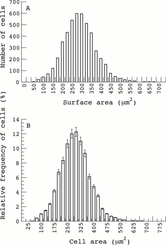
Figure 2 .

Group averaged histogram of endothelial cell areas. The area values were grouped into 0-25, 26-50, etc. and the entire data set pooled (A) or the average values within each group calculated and the mean (SEM) for each interval presented (B). In either case, the overall distribution of areas is clearly Gaussian.
Figure 3 .

Tracing overlay of the endothelial mosaic from Figure 1. The six sided cells are so indicated with the overall percentage of six sided cells being 61%, compared with the group averaged value of 66.6% (see Results). Bar = 0.05 mm.
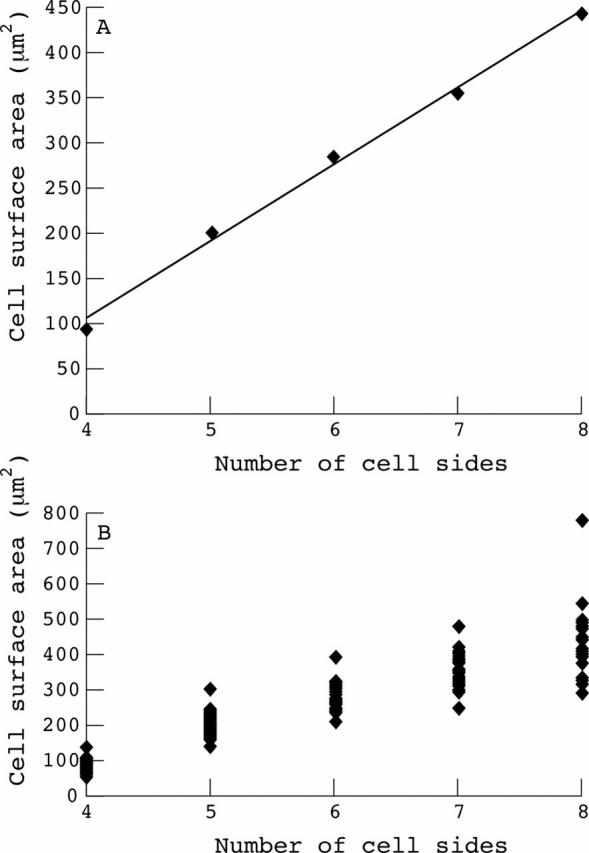
Figure 4 .

Area side relations for corneal endothelial cells. The group averaged mean value for the area of each cell type (four, five, six sided, etc) is plotted against the number of cell sides (A) or the mean values from each data set shown in the scatter plot (B). The linear regression shown in (A) is statistically significant (p <0.01; r2 value of 0.993), while the χ2 value across groups in (B) is highly significant at 142 (4 dF, p <0.01).
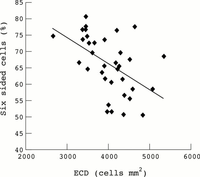
Figure 5 .
Relation between ECD and the percentage of six sided cells in the endothelium. The linear regression is statistically significant (p <0.01; r=0.492).
Selected References
These references are in PubMed. This may not be the complete list of references from this article.
- Bahn C. F., Glassman R. M., MacCallum D. K., Lillie J. H., Meyer R. F., Robinson B. J., Rich N. M. Postnatal development of corneal endothelium. Invest Ophthalmol Vis Sci. 1986 Jan;27(1):44–51. [PubMed] [Google Scholar]
- Bigar F. Specular microscopy of the corneal endothelium. Optical solutions and clinical results. Dev Ophthalmol. 1982;6:1–94. [PubMed] [Google Scholar]
- Bigar F. Specular microscopy of the corneal endothelium. Optical solutions and clinical results. Dev Ophthalmol. 1982;6:1–94. [PubMed] [Google Scholar]
- Blatt H. L., Rao G. N., Aquavella J. V. Endothelial cell density in relation to morphology. Invest Ophthalmol Vis Sci. 1979 Aug;18(8):856–859. [PubMed] [Google Scholar]
- Bourne W. M., Enoch J. M. Some optical principles of the clinical specular microscope. Invest Ophthalmol. 1976 Jan;15(1):29–32. [PubMed] [Google Scholar]
- Bourne W. M., Kaufman H. E. Specular microscopy of human corneal endothelium in vivo. Am J Ophthalmol. 1976 Mar;81(3):319–323. doi: 10.1016/0002-9394(76)90247-6. [DOI] [PubMed] [Google Scholar]
- Bourne W. M., Kaufman H. E. The endothelium of clear corneal transplants. Arch Ophthalmol. 1976 Oct;94(10):1730–1732. doi: 10.1001/archopht.1976.03910040504008. [DOI] [PubMed] [Google Scholar]
- Carlson K. H., Bourne W. M., McLaren J. W., Brubaker R. F. Variations in human corneal endothelial cell morphology and permeability to fluorescein with age. Exp Eye Res. 1988 Jul;47(1):27–41. doi: 10.1016/0014-4835(88)90021-8. [DOI] [PubMed] [Google Scholar]
- Chan-Ling T., Curmi J. Changes in corneal endothelial morphology in cats as a function of age. Curr Eye Res. 1988 Apr;7(4):387–392. doi: 10.3109/02713688809031788. [DOI] [PubMed] [Google Scholar]
- Doughty M. J. Are there geometric determinants of cell area in rabbit and human corneal endothelial cell monolayers? Tissue Cell. 1998 Oct;30(5):537–544. doi: 10.1016/s0040-8166(98)80034-7. [DOI] [PubMed] [Google Scholar]
- Doughty M. J. Concerning the symmetry of the 'hexagonal' cells of the corneal endothelium. Exp Eye Res. 1992 Jul;55(1):145–154. doi: 10.1016/0014-4835(92)90102-x. [DOI] [PubMed] [Google Scholar]
- Doughty M. J., Müller A., Zaman M. L. Assessment of the reliability of human corneal endothelial cell-density estimates using a noncontact specular microscope. Cornea. 2000 Mar;19(2):148–158. doi: 10.1097/00003226-200003000-00006. [DOI] [PubMed] [Google Scholar]
- Doughty M. J. Prevalence of 'non-hexagonal' cells in the corneal endothelium of young Caucasian adults, and their inter-relationships. Ophthalmic Physiol Opt. 1998 Sep;18(5):415–422. [PubMed] [Google Scholar]
- Doughty M. J. Toward a quantitative analysis of corneal endothelial cell morphology: a review of techniques and their application. Optom Vis Sci. 1989 Sep;66(9):626–642. doi: 10.1097/00006324-198909000-00010. [DOI] [PubMed] [Google Scholar]
- Hartmann C., Kolb M., Knauer I., Konen W. Klinische Spiegelmikroskopie. Technik, Organisation und einfache Kleinrechner-Morphometrie. Klin Monbl Augenheilkd. 1985 Feb;186(2):96–104. doi: 10.1055/s-2008-1050883. [DOI] [PubMed] [Google Scholar]
- Hiles D. A., Biglan A. W., Fetherolf E. C. Central corneal endothelial cell counts in children. J Am Intraocul Implant Soc. 1979 Oct;5(4):292–300. doi: 10.1016/s0146-2776(79)80078-6. [DOI] [PubMed] [Google Scholar]
- Hoffer K. J., Kraff M. C. Normal endothelial cell count range. Ophthalmology. 1980 Sep;87(9):861–866. doi: 10.1016/s0161-6420(80)35149-x. [DOI] [PubMed] [Google Scholar]
- Hoskovcová-Krejcová H., Hoskovec P., Krejcí L., Rezek P. Klasifikace endoteliálních nálezů u kongenitálního glaukomu pocítacovou analýzou. Cesk Oftalmol. 1985 Jul;41(4):252–257. [PubMed] [Google Scholar]
- Laule A., Cable M. K., Hoffman C. E., Hanna C. Endothelial cell population changes of human cornea during life. Arch Ophthalmol. 1978 Nov;96(11):2031–2035. doi: 10.1001/archopht.1978.03910060419003. [DOI] [PubMed] [Google Scholar]
- Matsuda M., Shiozaki Y., Suda T., Inoue Y., Manabe R. [Chronological change of human corneal endothelial cell shape and its arrangement]. Nippon Ganka Gakkai Zasshi. 1982;86(11):1944–1951. [PubMed] [Google Scholar]
- McCarey B. E. Noncontact specular microscopy: a macrophotography technique and some endothelial cell findings. Ophthalmology. 1979 Oct;86(10):1848–1860. doi: 10.1016/s0161-6420(79)35337-4. [DOI] [PubMed] [Google Scholar]
- Murphy C., Alvarado J., Juster R., Maglio M. Prenatal and postnatal cellularity of the human corneal endothelium. A quantitative histologic study. Invest Ophthalmol Vis Sci. 1984 Mar;25(3):312–322. [PubMed] [Google Scholar]
- Nucci P., Brancato R., Mets M. B., Shevell S. K. Normal endothelial cell density range in childhood. Arch Ophthalmol. 1990 Feb;108(2):247–248. doi: 10.1001/archopht.1990.01070040099039. [DOI] [PubMed] [Google Scholar]
- Price N. C., Barbour D. J. Corneal endothelial cell density in twins. Br J Ophthalmol. 1981 Dec;65(12):812–814. doi: 10.1136/bjo.65.12.812. [DOI] [PMC free article] [PubMed] [Google Scholar]
- Schimmelpfennig B. Topographie altersbedingter Grössenveränderungen von Kornea-Endothelzellen. Klin Monbl Augenheilkd. 1984 May;184(5):353–356. doi: 10.1055/s-2008-1054488. [DOI] [PubMed] [Google Scholar]
- Sherrard E. S., Novakovic P., Speedwell L. Age-related changes of the corneal endothelium and stroma as seen in vivo by specular microscopy. Eye (Lond) 1987;1(Pt 2):197–203. doi: 10.1038/eye.1987.37. [DOI] [PubMed] [Google Scholar]
- Speedwell L., Novakovic P., Sherrard E. S., Taylor D. S. The infant corneal endothelium. Arch Ophthalmol. 1988 Jun;106(6):771–775. doi: 10.1001/archopht.1988.01060130841036. [DOI] [PubMed] [Google Scholar]
- Stefansson A., Müller O., Sundmacher R. Non-contact specular microscopy of the normal corneal endothelium. A statistical evaluation of morphometric parameters. Graefes Arch Clin Exp Ophthalmol. 1982;218(4):200–205. doi: 10.1007/BF02150095. [DOI] [PubMed] [Google Scholar]
- Suda T. Mosaic pattern changes in human corneal endothelium with age. Jpn J Ophthalmol. 1984;28(4):331–338. [PubMed] [Google Scholar]
- Tsukahara Y., Yamamoto M. [Postnatal development of corneal endothelial cells in normal children]. Nippon Ganka Gakkai Zasshi. 1989 Jul;93(7):763–768. [PubMed] [Google Scholar]
- Waring G. O., 3rd, Bourne W. M., Edelhauser H. F., Kenyon K. R. The corneal endothelium. Normal and pathologic structure and function. Ophthalmology. 1982 Jun;89(6):531–590. [PubMed] [Google Scholar]
- Williams K. K., Noe R. L., Grossniklaus H. E., Drews-Botsch C., Edelhauser H. F. Correlation of histologic corneal endothelial cell counts with specular microscopic cell density. Arch Ophthalmol. 1992 Aug;110(8):1146–1149. doi: 10.1001/archopht.1992.01080200126039. [DOI] [PubMed] [Google Scholar]
- Wulle K. G. Electron microscopy of the fetal development of the corneal endothelium and Descemet's membrane of the human eye. Invest Ophthalmol. 1972 Nov;11(11):897–904. [PubMed] [Google Scholar]


