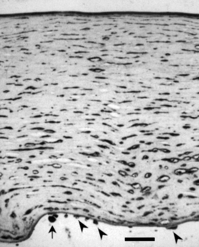
Figure 1 .

Case 1. The cornea is devoid of epithelium and the posterior surface is bowed as a result of oedema. Virtually all keratocytes are strongly positive for HSV-1, as are the remaining endothelial cells (arrowheads). Bowman's layer and Descemet's membrane are diffusely positive, possibly reflecting viral load in this tissue. A pigment-containing phagocyte (arrow) is present on the posterior surface of the cornea. (Immunohistochemistry, anti-HSV-1, DAB with haematoxylin counterstain. Scale bar = 100 µm).
