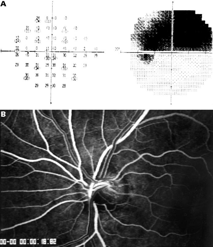
Figure 2 .
(A) Asymmetric hemifield visual field loss of the superior visual field (Humphrey 24-2). (B) Corresponding fluorescein angiogram with ischaemia of the inferior portion of the optic disc (AVP of affected inferior vessel formation: 3.6 seconds; AVP of less affected superior vessel formation: 2.23 seconds). Note the visible different fluorescein intensity of the laminar stream of the dye in the veins superiorly and inferiorly.

