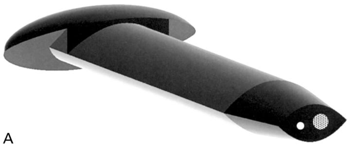
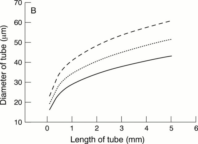
Figure 6 .
(A) This is one embodiment of a novel glaucoma drainage device (patent applied for) incorporating a small diameter hole (white circle) to provide the required fistular resistance and a larger diameter hole (dotted circle) temporarily occluded by a thin ablatable membrane. To give the lowest final IOP, resistance is bypassed by ablating this thin membrane using a YAG laser delivered through a gonioscope once a mature bleb is established. The external cross sectional shape of this implant is designed to bear evenly on the internal aspect of a standard microvitreoretinal (MVR) slit incision, so as to eliminate external leakage after placement. (B) A schematic drawing of the glaucoma drainage device in situ. A limbal slit incision was made parallel to the iris plane using a standard 1.19 mm width MVR blade to enter the anterior chamber. The occluded glaucoma drainage device was inserted through the wound, and sutured with 10/0 nylon.


