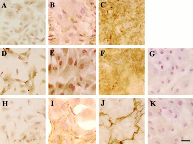
Figure 4 .
Immunolocalisation of LTBP-1, β1-LAP, and fibrillin-1 in the extracellular matrix accumulation in bovine lens epithelial cell cultures. In sparse, preconfluent, cultures at day 3, almost no immunoreactivity for LTBP-1 was detected in LECs (A). In early confluence, a small amount of LTBP-1 was detected extracellularly in a very fine fibrillar pattern (B), while in mature confluent cultures at day 14, immunoreactivity for LTBP-1 appeared to form an organised network throughout the culture (C). Immunoreactivity for β1-LAP was detected in the cells of a sparsely cellular culture (D) and an early confluent culture (E). The mature confluent culture of LECs exhibited extracellular deposition of β1-LAP (F). In sparsely cellular cultures, LECs did not express fibrillin-1 protein (H). In early confluent cultures at day 10, fibrillin-1 was detected in a fine fibrillar pattern in fibroblast ECM; the cells themselves were unstained (I). Mature confluent LEC cultures showed a condensed fibrillin-1 structure (J). No specific immunoreactivity was observed in negative controls with anti-goat IgG (G) or anti-rabbit IgG but no primary antibody (K). LTBP-1 = large transforming growth factor β binding protein-1; β1-LAP = β1-latency associated peptide; LEC = lens epithelial cells. Indirect immunostaining; bar = 50 µm.

