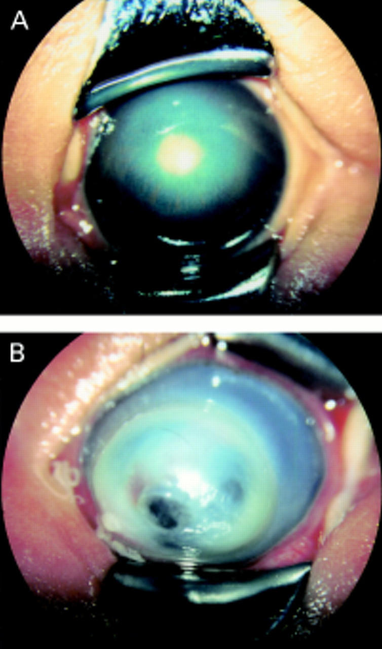
Figure 1 .

(A) A photograph of anterior segment of case 1. Central corneal opacity, elongated ciliary processes, and whitish mass behind the lens are observed. (B) A photograph of anterior segment of case 2. The cornea is noticeably protruding and densely opaque overall. These findings correspond to anterior staphyloma.
