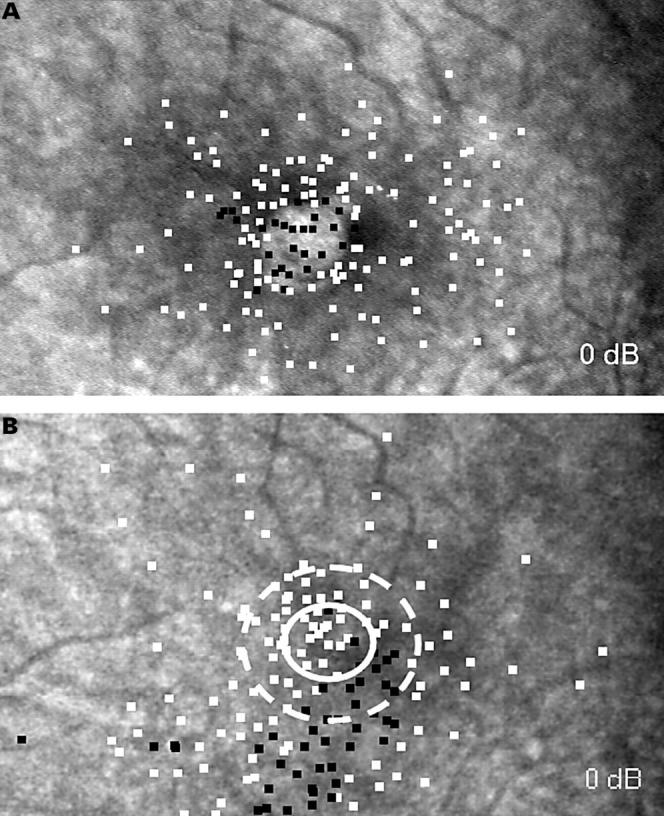
Figure 1 .

(A) Preoperative and postoperative (B) microperimetry of a left eye. White squares represent stimuli that were seen by the patient, black squares represent relative (12 dB) or deep (0 dB) scotomata. The white circle marks the area of the preoperative macular defect, the broken circle the area of the preoperative neurosensory detachment. Postoperatively a large deep scotoma inferior to the closed macular hole can be seen in an area that was tested normally before surgery. The scotoma had the shape of a nerve fibre bundle defect.
