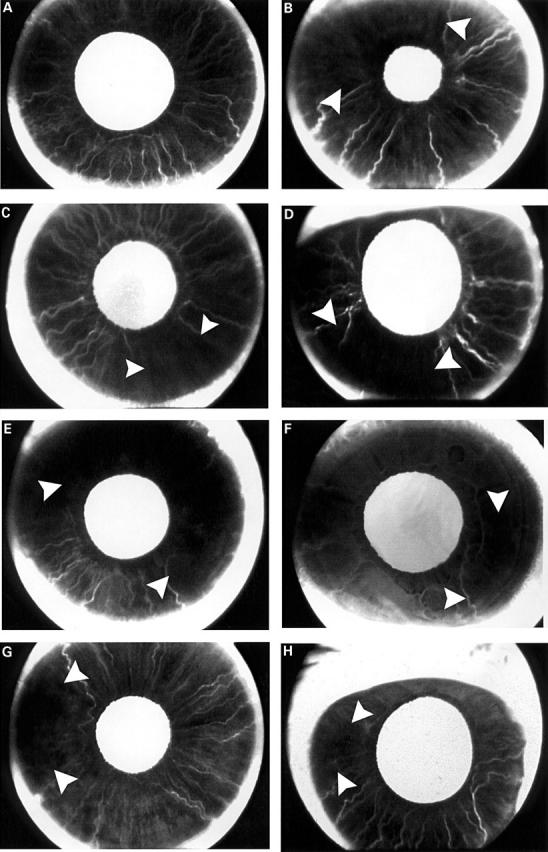
Figure 1 .

(A) Normal dark brown iris ICGA of the right eye in a 49 year old man. Late frame at 46 seconds showed complete filling of the iris. (B) ICGA of the right eye in a 68 year old man. One day after primary SR surgery. Superior and temporal sectors (arrowheads) filled out of phase by 34 seconds. (C) ICGA of the left eye in a 52 year old man. One day after primary IR surgery. The nasal, superior, and temporal sectors started to fill first which was followed by a delay in filling in inferior quadrant (arrowheads) by 40.2 seconds. (D) ICGA of the right eye in a 55 year old woman. One day after primary SR and IR surgery. Superior and inferior temporal sectors (arrowheads) showed delay in perfusion by 25 seconds. (E) ICGA of the left eye in a 59 year old man. One day after primary SR and MR surgery. Persistent superior sector delay (arrowheads) by 6 minutes; no nasal sector delay in filling. (F) ICGA of the left eye (previous 5 month history of LR and MR surgery) in a 58 year old woman. One day after IR and LR surgery. Inferior and temporal sectors filled out of phase (arrowheads). Nasal sector perfusion was normal. (G) ICGA of the right eye (previous 3 year history of MR surgery) in a 81 year old man. Preoperative ICGA showed persistent temporal sector delay at late frames. Change of surgical plan and limited surgery to LR only in this eye. One day after LR surgery. Temporal sector delay (arrowheads) persisted at 38.8 seconds. (H) ICGA of the right eye (previous 18 month history of SR and IR surgery) in a 15 year old girl. One day after MR and LR surgery. Temporal sector delay (arrowheads) in filling of 27.9 seconds and nasal sector was well perfused at early frames because of the MR anterior ciliary artery preservation technique 2 months after surgery.
