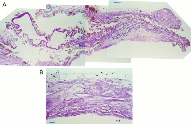
Figure 2 .
Specimen corresponding to a recent tear (case 5). (A) At one edge of the specimen intra-Bruch's fibrovascular tissue is recognised, more centrally to it rolled up RPE and diffuse drusen are seen that are only partially covered by fibrovascular tissue and amorphous debris on their retinal side. (B) Towards the opposite edge, rather fibrous fibrovascular membrane is observed denuded of RPE and diffuse drusen but covered by a thin layer of amorphous debris. Periodic acid Schiff, bar = 50 µm in (A) and 25 µm in (B).

