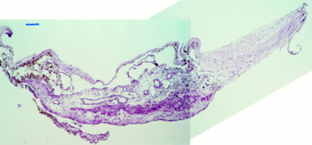
Figure 4 .
Specimen corresponding to a less recent tear (case 8). Rolled up RPE and diffuse drusen, which are covered by a thin fibrocellular membrane on their retinal side, are readily recognised. On their choroidal side fibrovascular tissue is observed with large bore vessels and a moderately strong inflammatory cell infiltration. Adjacent to this part of the specimen, fibrovascular tissue denuded of RPE and diffuse drusen but covered by a thin layer of amorphous debris is found. At one edge of the specimen, RPE, diffuse drusen, and a thin layer of intra-Bruch's fibrovascular tissue are found that has, however, turned over as shown on serial sectioning. At the opposite edge, RPE, diffuse drusen and a thin layer of intra-Bruch's fibrovascular tissue is found that has also turned over but does not physically make contact with the edge of the section itself at this level. Periodic acid Schiff, bar = 100 µm.

