
Figure 3 .
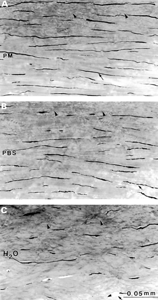
Light micrographs of the most anterior stroma in (A) post mortem (PM); (B) phosphate buffered saline (PBS), and (C) deionised water (H2O). In (A) and (B) more keratocyte profiles are present (arrows) than in (C). The undulated collagen bundles (arrowheads) in (A) and (B) are similar; (C) is different and characterised by irregularly arranged grey structures.