
Figure 1 .
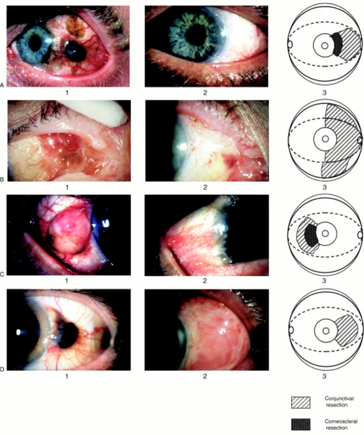
(A1) Preoperative view of a nodular, deeply pigmented conjunctival malignant melanoma arising in the context of primary acquired melanosis and a naevus in the temporal (epibulbar) quadrants of the left eye in a 49 year old man (case 1). (A2) Excellent cosmetic and functional result at 8 weeks after conjunctival and corneoscleral excisional surgery, corneoscleral grafting combined with amniotic membrane grafting (AMT), followed by cryotherapy and a topical mitomycin course (case 1). (A3) Schematic drawing depicting the areas of conjunctival and corneoscleral resection in case 1. (B1) Preoperative view of an irregular, partly pigmented elevated malignant melanoma involving the medial area of epibulbar conjunctiva, the plica, and the caruncle of the right eye in a 70 year old woman. The tumour originates from flat, lightly pigmented melanosis, extending into the medial part of the superior and inferior fornix and palpebral conjunctiva, and the lower lid margin (case 2). (B2) At 3 weeks after excisional surgery and AMT, while the patient was still using topical steroids, there was no sign of symblepharon formation. In the following weeks, when the steroids were tapered down, a fibrotic reaction underneath the amniotic membrane was noted (case 2). (B3) Schematic drawing depicting the areas of excised conjunctiva in case 2. (C1) Preoperative view of a nodular, amelanotic malignant melanoma at the limbus of the right eye in a 54 year old Asian man (case 3). (C2) No sign of local recurrence at 2 years following excisional surgery, cryotherapy, and AMT. Note the pterygium-like reaction of the transplanted area, with mild fibrosis and hyperaemia (case 3). (C3) Schematic drawing depicting the areas of corneoscleral and conjunctival resection in case 3. (D1) Preoperative view of a nodular, darkly pigmented, limbal malignant melanoma of the left eye in a 64 year old man (case 4). (D2) Satisfactory cosmetic and functional result at 4 weeks following tumour excision and AMT. The oedema and hyperaemia of the graft area is subsiding (case 4). (D3) Schematic drawing depicting the areas of conjunctival resection in case 4.