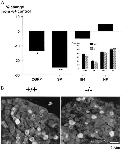
Figure 1.
(A) The percentage of cells histochemically expressing the markers CGRP, substance P (SP), IB4, and neurofilament (NF) in adult wild-type and mutant DRG. (Inset) Graph represents absolute values rather than percentage change for each marker. Significant deficits were observed in the percentage of CGRP- and substance P- expressing neurons, whereas the percentages of cells expressing IB4 and NF were unchanged (t test; *, P < 0.05; **, P < 0.01, n = 6). It should be noted that these markers are not completely exclusive of each other and in some cases may colocalize in single neurons. (B) Representative photomicrograph demonstrating fewer substance P-positive cells in the DRG from mutant (Right) as compared with wild-type (Left) animals.

