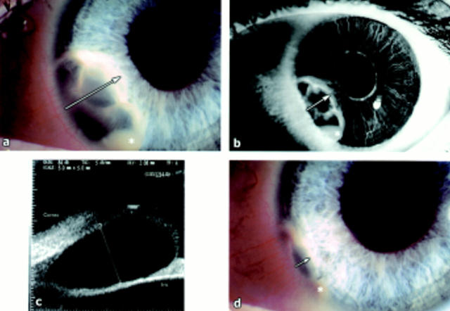
Figure 1 .
(a) Iris stromal cyst at first presentation with the asterisk representing the pseudohypopyon and the arrow indicating the radial extent of the cyst. (b) Anterior segment fluorescein angiography showing leakage with the asterisk representing the pseudohypopyon and the arrow indicating the radial extent of the cyst. The white spot at 4 o'clock is an artefact. (c) Ultrasound biomicroscopy of the cyst showing origin from superficial stroma. The cornea and iris have been labelled and the cursor indicates the anteroposterior extent of the cyst. (d) Resolving cyst after YAG laser treatment with the asterisk representing the pseudohypopyon and the arrow indicating the radial extent of the cyst.

