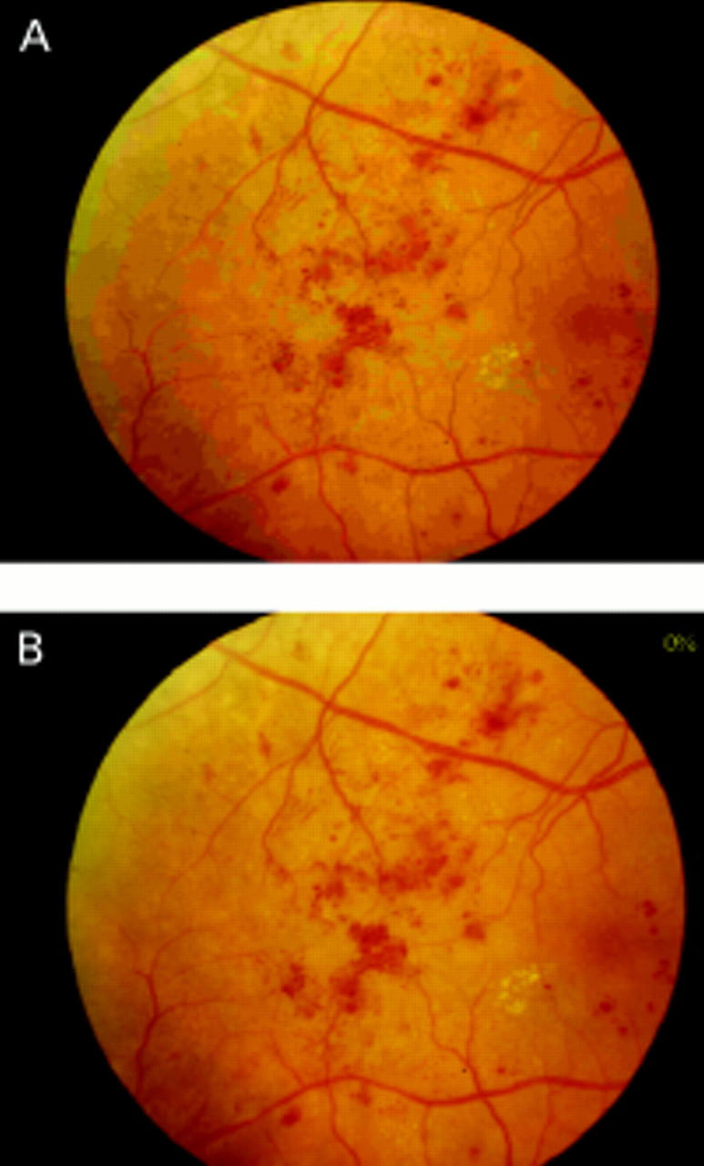
Figure 1 .

Image compression showing the effect of image compression on an image with proliferative retinopathy. (A) 90% compression: note colour blocking, increased granularity, with loss of intraretinal microvascular abnormalities, microaneurysms, and neovascularisation. (B) 0% compression: showing good retinal detail, fine details clear on image.
