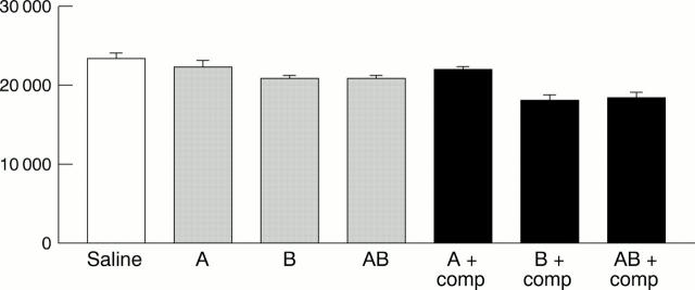
Figure 2 .
Shows live cells remaining after blood group B corneal epithelial cells were incubated with the various antisera, with and without complement. Significant lysis was observed with anti-B + complement and anti-AB + complement. (A = anti-A antibody, B = anti-B antibody, AB = anti-AB antibody, comp = complement.) The Y axis represents scintillation counts (0.02 minutes/well) of live cells. Error bars represent SEM.

