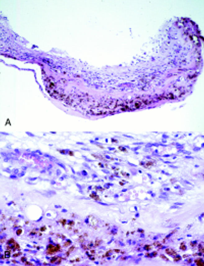
Figure 3 .

(A) General view of CNV after PDT represents hyaline masses between retinal pigment epithelium and fibrovascular tissue with severe oedema (haematoxylin and eosin staining, ×100). (B) The hyalinised masses in the CNV were partly surrounded by small red cell containing vessels partly invading the CNV (haematoxylin and eosin ×400).
