
Figure 1 .
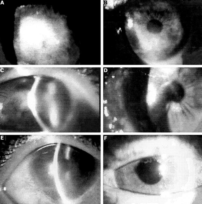
Preoperative and postoperative photographic sequence of three patients undergoing ILT. (A) Patient 1 with 4+ conjunctival hyperaemia, central epithelial defect, corneal oedema, neovascularisation, and BCVA of hand movements. (B) Same patient 11 months after ILT. Stable ocular surface and BCVA of 20/40. (C) Patient 2 presenting with diffuse conjunctival hyperaemia and corneal opacity with epithelial oedema, neovascularisation, and severe stromal oedema. BCVA was hand movements. (D) Same patient 10 months after ILT. BCVA was 20/60. (E) Patient 3 presenting epithelial defect, corneal oedema, and stromal opacity compromising the third inferior section of the cornea. BCVA was 20/80. (F) Nine months after ILT BCVA was 20/20.