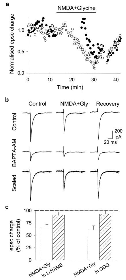
Figure 3.
Mechanism of the NMDA-induced depression of synaptic transmission. (a) Effect of application of 30 μM NMDA + 10 μM glycine on EPSCs evoked by two different stimulation electrodes. Stimulation 1 (○) was applied every 20 s during the whole experiment. Stimulation 2 (●) was applied at the same frequency but was stopped immediately before agonist application and resumed 10 min later. EPSC charge is normalized to the mean value of the period preceding agonist application (stimulation 1: 4.2 pC; stimulation 2: 5.7 pC). Note that as few as 30 action potentials in the presence of NMDA are sufficient to induce a 50% decrease of the PF EPSC. (b) Reducing glutamate release by incubating the slice in BAPTA-AM (50 μM, 5 min) reduced the PF-PC EPSC but did not modify the effect of NMDA. When the responses recorded after BAPTA-AM treatment and in the absence of NMDA were scaled up to the amplitude of the control EPSCs (“scaled”) (control responses: solid line; BAPTA-AM: dotted line), the responses recorded in the presence of NMDA scaled by the same factor were also superimposable. (c) Effect of 30 μM NMDA + 10 μM glycine application on EPSC charge in control conditions (empty bars) or in the presence of l-NAME (1 mM) or ODQ (1 μM) (hatched bars). Bars represent means ± SEM of three experiments.

