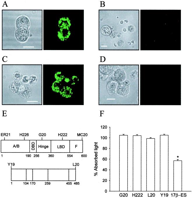
Figure 4.
(A) Anti-ERα antibody H222 stains the cytoplasm of permeabilized β cells in primary culture. (B) Absence of staining at the plasma membrane of nonpermeabilized β cells with anti-ERα antibody H222. These cells were from the same culture as the cells in A. (C) Anti-ERβ antibody Y19 stains the cytoplasm of permeabilized β cells in primary culture. (D) Lack of staining of Y19 antibody at the membrane of nonpermeabilized cells. These cells were from the same culture as those in C. Fluorescence pictures are on the right and their corresponding transmission pictures are on the left. Results are representative of three different duplicated experiments. (E) Epitopes of the intracellular ERα (upper map of the peptide) and ERβ (lower map of the peptide) by available anti-ERα and anti-ERβ antibodies. Epitopes are located by the antibodies named above. (F) Impeded binding ligand assay. E-HRP binding was not blocked either by anti-ERα antibodies H222 and G20 or by anti-ERβ antibodies Y19 and L20. Data are from three duplicated experiments, expressed as mean ± SEM, *, P < 10-5 Student's t test. (Scale bars are 10 μm.)

