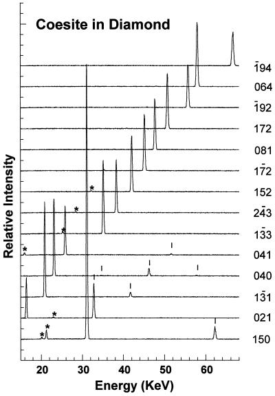
Figure 3.
Single-crystal energy dispersive x-ray diffraction patterns measured on the small coesite inclusion, collected at fixed 2θ = 10.002° (E d = 70.094 keV-Å; 1 eV = 1.602 × 10⁻¹⁹ J) and room temperature. Reflections of 14 different h k l, randomly picked from a total of 56, are shown. Each reflection was obtained at a distinct χ, ω diffraction direction dictated by the orientation matrix of the single crystal. Short vertical ticks mark the overtones (nh nk nl), and the asterisks mark Ge detector escape peaks.

