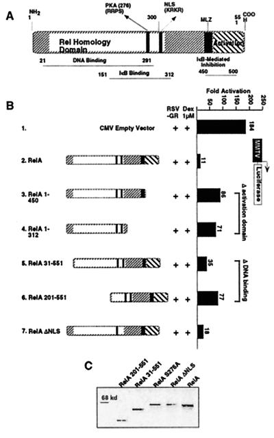
Figure 2.
Mapping the domain of RelA involved in GR repression. (A) Schematic of RelA. (B) RelA cross-coupling domains. CV1 cells (48-well plates) were transiently cotransfected with 120 ng of MMTV-Luc reporter construct, 75 ng of CMXβgal, 37.5 ng of RSVGR, and CMV empty vector or CMVRelA mutants at 3:1 M ratio with GR, as indicated. Cells were stimulated with Dex at 1 μM for 10 h before the assay. The y axis shows activation as measured by MMTV-driven Luc activity conducted in quadruplicate assays. (C) Expression levels of RelA proteins. Western blot analysis of RelA wild-type and mutant proteins.

