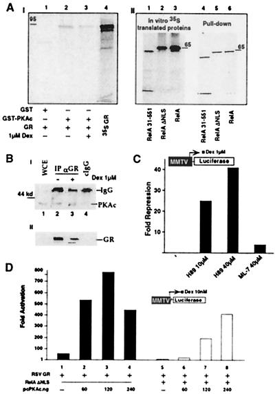
Figure 5.
PKAc is associated with GR and potentiates GR activity. (A) In vitro association of PKAc subunit and GR (95 kDa). (AI) The 35S-labeled GR protein was incubated with GST-PKAc or GST alone on glutathione-agarose beads, in the absence or presence of 1 μM Dex as indicated. The bound proteins were analyzed by SDS/PAGE and fluorography (lanes 1–3). Input was included in lane 4. (AII) The same conditions as in AI but using 35S-labeled RelA wild-type and mutants. Input proteins (lanes 1–3) and the pull-down proteins (lanes 4–6) are indicated. (B) In vivo association of PKAc and GR wild-type protein. (BI) Human 293 cells were cotransfected with CMXGR and pcPKAc expression vectors. Cells were mock-treated (lane 2) or activated with 1 μM Dex for 12 h (lane 3) and harvested 48 h after transfection. Cell lysates were immunoprecipitated with 5 μl of GR-135 polyclonal antibody (lanes 2 and 3) or with the same amount of normal rabbit IgG as a control (lane 4). Immunoprecipitates then were analyzed by Western blot with PKAc-specific antibody. WCE were loaded in parallel with the immunoprecipitates to show comigration (lane 1). (BII) An aliquot of the WCE was analyzed directly with Western blot by using GR-specific antibody. (C) H-89, a PKAc-specific inhibitor represses GR activity. CV-1 cells were transfected with MMTV-Luc, CMXβgal, and RSVGR as in Fig. 2B. After transfection cells were stimulated for 4 h with 1 μM Dex, with or without H-89 or ML-7, at the indicated concentrations. Cellular extracts were used to measure Luc activity. The y axis shows arbitrary unit of repression as measured by MMTV-driven Luc activity. The histogram is representative of at least three independent experiments. (D) PKAc potentiates GR transcription and reverses RelA repression of GR. CV1 cells were cotransfected with 120 ng of MMTV-Luc reporter construct, 75 ng of CMXβgal, 33 ng of RSVGR, CMV empty vector or CMVRelA ΔNLS at 120 ng, and increasing concentration of PKAc expression vector, as indicated. Cells were stimulated with Dex at 10 nM for 10 h before the assay. All of the transfections were normalized for the total amount of expression vectors, i.e., CMV empty vector. The y axis shows arbitrary unit of activation as measured by MMTV-driven Luc activity.

