
Figure 6 .
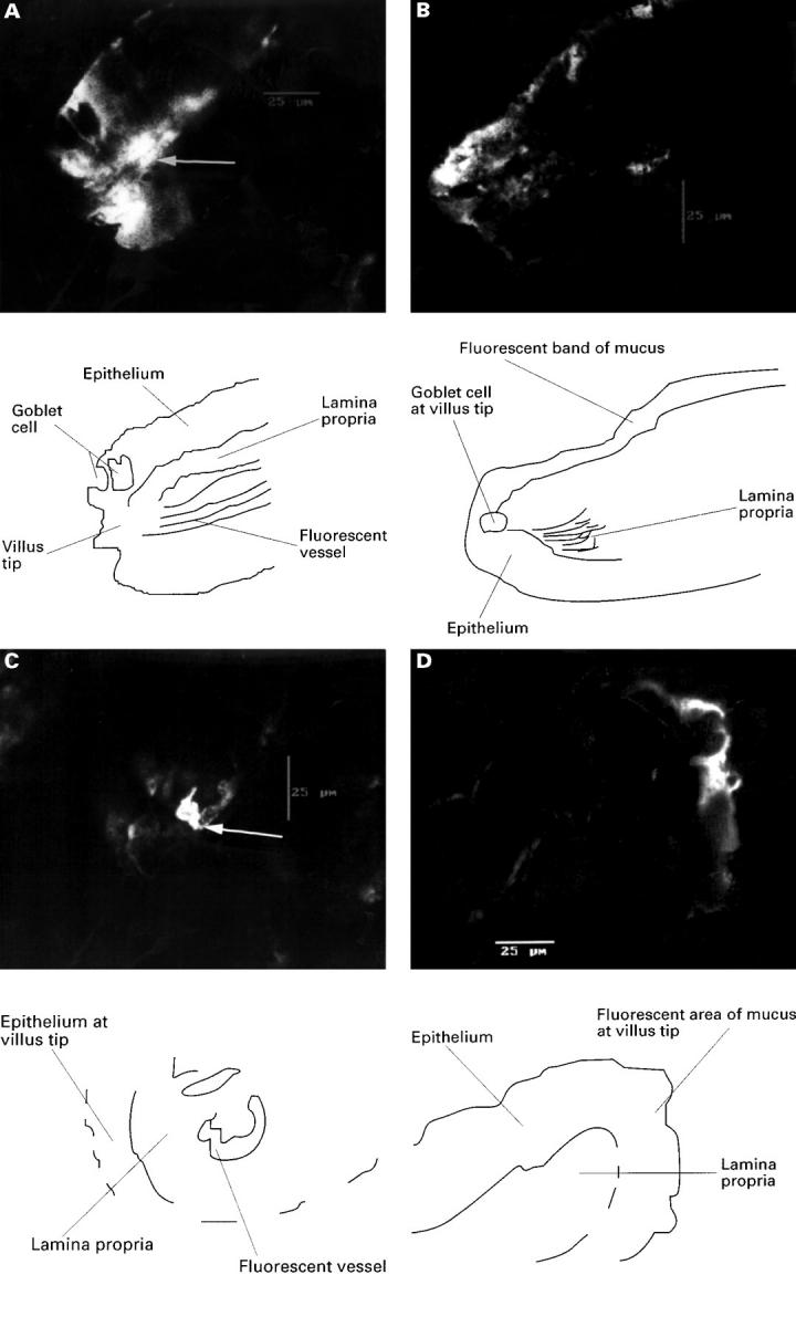
(A) An isolated villus from rat jejunum after combined indomethacin, taken from the left half of the villus which showed stasis and vessel hyperfluorescence (arrow), at which point the villus was perfuse fixed; the area of fluoresence to the left of this vessel is epithelium; original magnification ×600. This is more clearly seen in the line drawing. (B) Image from other right side of the villus which had normal flow and no fluorescence except for surface mucus; original magnification ×600. This is more clearly shown in the line drawing. (C) Transverse confocal image of an isolated villus from rat jejunum after combined luminal (100 µg/ml, 2.8 × 10⁻⁴M) and intravenous (15 mg/kg) indomethacin, and perfusion fixed as blood flow slowed at 30 minutes. Image is taken from the right side of the villus which exhibited slowing of flow and hyperfluorescence of the arcade artery (arrow), which is surrounded by epithelium showing slight fluoresence due to surface mucus; original magnification ×600. This is more clearly shown in the line drawing. (D) The left side of this villus which maintained normal flow. The only fluorescence again comes from surface mucus on the epithelium; original magnification ×600. This is more clearly shown in the line drawing.