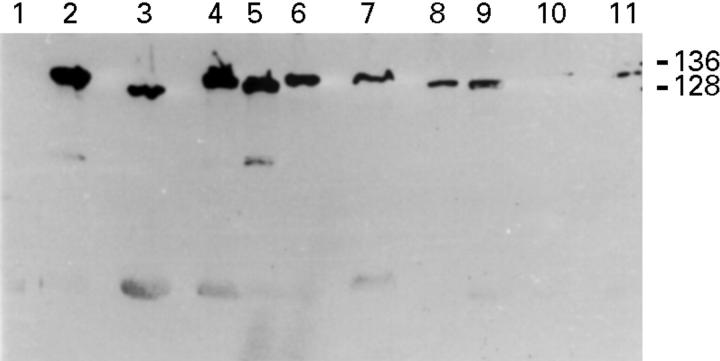
Figure 2 .
Western blots showing , in three cases (lanes 1-3, 4-6, and 7-9), the simultaneous presence of H pylori strains with different CagA molecular mass in different areas of single patients. The strain in lane 1 was cagA−. The strains in lanes 10 and 11 were two cagA+ H pylori strains of similar CagA molecular mass isolated from two different gastric areas of a patient. The numbers on the right are the molecular masses in kDa of H pylori strains CCUG 17874 and G39.

