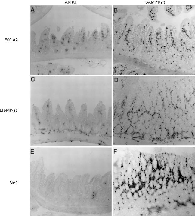
Figure 3 .
Immunohistochemical staining of the ileal mucosa. A cryostat section from IBD SAMP1/Yit and age matched control AKR/J mice was stained with monoclonal antibodies 500-A2, ER-MP-23, and Gr-1 that recognise CD3-ε, tissue macrophages, and granulocytes, respectively. CD3-ε positive cells, tissue macrophages, and granulocyes were more abundant in the IBD lesion in SAMP1/Yit (B,D,F) than in age matched control AKR/J mice (A,C,E). Original magnification, ×100.

