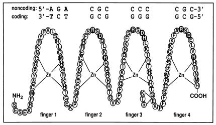
Figure 1.
Schematic representation of the WT1 zinc-finger domain (fingers 1–4; residues 318–438) with the core DNA recognition element showing the “antiparallel” orientation in which the complex is formed (the 5′ end of the coding strand of the DNA is aligned with the C terminus of the WT1 protein). The circles representing the putative DNA base-contacting residues (14, 23, 35) are shaded black. Base-contacting residues in finger 1 are unknown, and those in finger 4 occur only in the −KTS isoform (see text). The KTS insertion site between fingers 3 and 4 is indicated.

