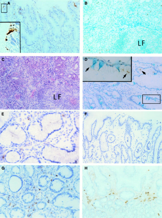
Figure 4 .

Immunohistochemical analysis of H pylori urease specific humoral responses. (A) Localisation of H pylori and H pylori urease in the stomach of a patient with grade III gastritis (enzyme immunohistochemistry with monoclonal H pylori urease specific antibody; Meyer's haematoxylin, original magnification ×160); a higher maginification (×800) of the boxed area indicated in the left upper corner is shown in the left lower corner. (B) Distribution of H pylori urease specific antibody producing B lymphocytes (plasma cells) in the same tissue (enzyme immunohistochemistry with purified H pylori urease and monoclonal H pylori urease specific antibody; methyl green, original magnification ×150); LF, lymphoid follicle. (C) Localisation of mononuclear cells in the same tissue (haematoxylin and eosin, original magnification ×150). (D) Localisation of H pylori and H pylori urease indicated by arrows in the stomach of a patient with grade II gastritis (enzyme immunohistochemistry with monoclonal H pylori urease specific antibody; Alcian blue, original magnification ×160); a higher magnification (×640) of the boxed area indicated in the right lower corner is shown in the left upper corner. (E) Distribution of H pylori urease specific B cells in the stomach from the same patient (enzyme immunohistochemistry with purified H pylori urease and monoclonal H pylori urease specific antibody; Meyer's haematoxylin, original magnification ×250). (F) Distribution of H pylori urease specific plasma cells in the stomach of a patient with grade I gastritis (enzyme immunohistochemistry with purified H pylori urease and monoclonal H pylori urease specific antibody; Meyer's haematoxylin, original magnification ×125). (G) Detection of H pylori urease specific B cells in the duodenal bulb from the same patient with grade I gastritis bearing duodenitis (enzyme immunohistochemistry with purified H pylori urease and monoclonal H pylori urease specific antibody; Meyer's haematoxylin, original magnification ×200). (H) Distribution of IgA producing B cells in the stomach of the same patient (enzyme immunohistochemistry with antihuman IgA antibody, original magnification ×132).
