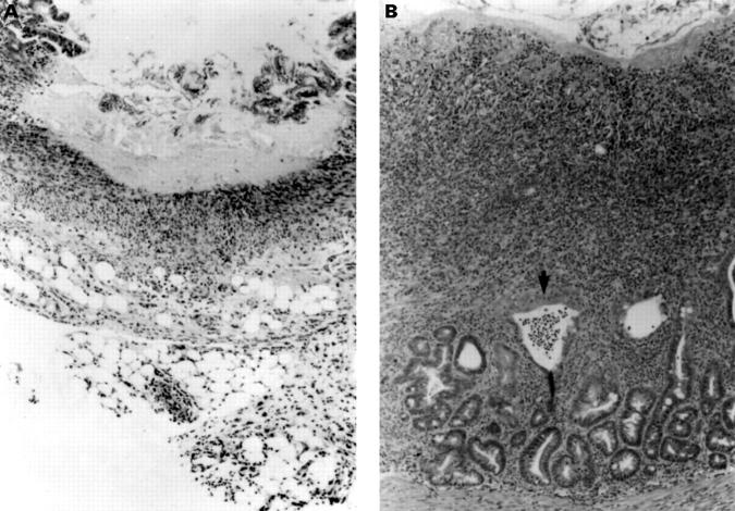
Figure 1 .
Histological evidence of enteritis induced by indomethacin in female Lewis rats. (A) At day 2, after subcutaneous injection of 7.5 mg/kg indomethacin for two days, a large ulcer with relatively bland necrosis of the entire mucosa is seen with severe serosal and mesenteric inflammation with neutrophils and macrophages (original magnification ×100). (B) At day 14, there is an extensive ulcer with an active mucosal exudate. A crypt abscess is visible (arrow) and glandular dysplasia with depletion of goblet cells. Transmural inflammation with thickening of the submucosa is evident. The infiltrate consists of both mononuclear cells and neutrophils with fibroblast proliferation and smooth muscle hypertrophy. Collagen indicative of early fibrosis is present (original magnification ×100).

