
Figure 6 .
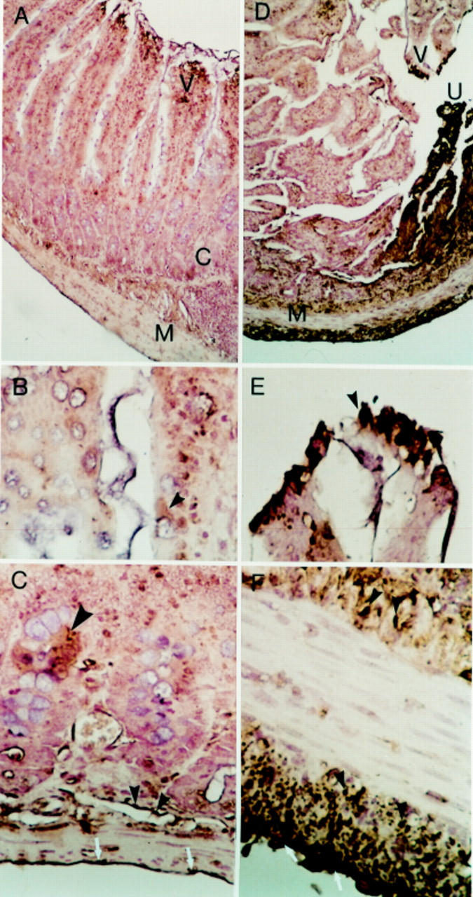
Immunolocalisation of the kinin B2 receptor in rat intestine on day 2. A-C: Rat normal control jejunum (buffer treated). (A) Some focal staining positive for B2 receptor in the villi—V, occasionally in the muscularis mucosa below the crypts—C, in the muscular layer—M, and in the serosa. Original magnification ×100. (B) Intestinal villi. Focal weak immunostaining in the surface of occasional epithelial cells and in cytoplasm (arrow). Original magnification ×400. (C) Some focal immunostaining, cluster of epithelial cells in crypts (large arrow), in the muscular layers (small solid arrows), and in serosal surface (white arrows). Original magnification ×400. D-F: Rat inflamed intestine (day 2 after injection of indomethacin). (D) Focal immunostaining in the villi—V, especially near the area of ulceration - U, in the internal and external muscular layer—M, and in the serosa. Original magnification ×100. (E) Villus near area of ulceration showing strong staining in the epithelial, probably absorptive cells (arrow). Original magnification ×400. (F) Prominent immunostaining in the smooth muscle cells of the muscularis mucosa (solid arrows) and in the external muscular layer (solid arrows). On the serosal surface, staining is also present (white arrows). Original magnification ×400.