
Figure 1 .
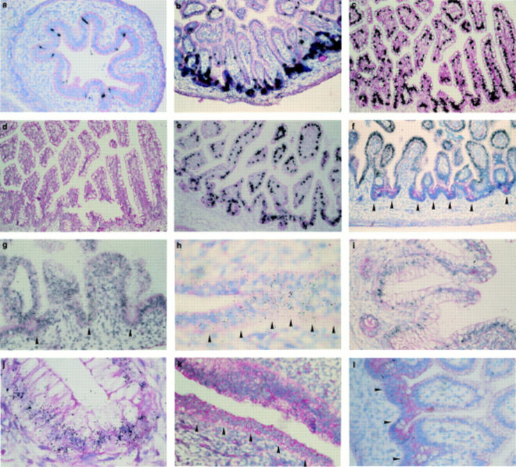
In situ hybridisation for mucin gene mRNAs in embryonic and fetal intestine. (a) Small intestine at 10 weeks gestation with MUC2 probe: the signal is located predominantly between the primordial villi (methyl green pyronin counterstain; original magnification ×200). (b) Small intestine at 12 weeks' gestation with MUC2 probe: the signal is located predominantly within immature crypts of Lieberkühn (methyl green pyronin counterstain; original magnification ×200). (c, d) Ileum at 23 weeks' gestation: (c) with MUC2 probe: signal is stronger in the crypts, but the majority of villous goblet cells are labelled; (d) with MUC2 probe and an excess of cold unlabelled MUC2 oligonucleotide (negative control): hybridisation is negative (methyl green pyronin counterstain; original magnification ×200). (e) Ileum at 26 weeks' gestation with MUC2 probe: signal is distributed in goblet cells both on villi and in crypts (methyl green pyronin counterstain; original magnification ×200). (f) Small intestine at 12 weeks' gestation with MUC3 probe: continuous and homogeneous labelling along the villous epithelium, both in goblet and absorptive cells; no labelling present in crypts (arrows) (methyl green pyronin counterstain; original magnification ×400). (g) Small intestine at 14.1 weeks' gestation with MUC3 probe: continuous and homogeneous labelling along the villous epithelium, both in goblet and absorptive cells; weak labelling is also present in crypts (arrows) (methyl green pyronin counterstain; original magnification ×500). (h) Anterior portion of primitive gut at 6.5 weeks' gestation with MUC4 probe: continuous labelling along epithelium (methyl green pyronin counterstain; original magnification ×1000). (i, j) Colon at 23 weeks' gestation with MUC4 probe: signal is located in the perinuclear region of goblet cells (methyl green pyronin counterstain; original magnification: (i) ×400; (j) ×1000). (k) Middle portion of primitive gut at 8 weeks' gestation with MUC5AC probe: continuous labelling along the undifferentiated epithelium (methyl green pyronin counterstain; original magnification ×500). (l) Ileum at 12 weeks' gestation with MUC5AC probe: labelling is limited to clusters of epithelial cells both on villi and in crypts (methyl green pyronin counterstain; original magnification ×500).