Figure 1 .

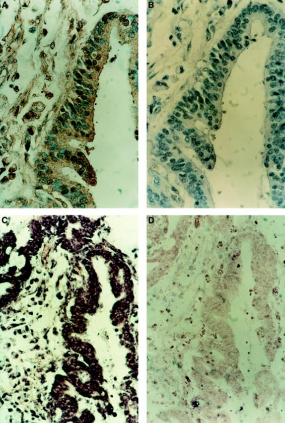
Expression of Fas ligand (FasL) in human gastric adenocarcinomas. Immunoperoxidase staining using a FasL specific rabbit polyclonal IgG was performed on paraffin wax embedded gastric carcinoma sections. Slides were counterstained with haematoxylin. (A) FasL positive immunohistochemical staining (brown) is shown in a representative gastric adenocarcinoma. (B) As a control for specificity of antibody detection, the FasL immunising peptide was included during primary antibody incubation. Competitive displacement of staining by the immunising peptide confirms FasL specificity. Tumour sections sequential to those shown in (A) and (B) were used to detect FasL mRNA by in situ hybridisation, using a digoxigenin labelled FasL specific riboprobe. (C) Positive purple hybridisation signal was obtained from the tumour area that stained immunohistochemically positive for FasL protein. (D) In a control hybridisation, a tenfold excess of unlabelled probe caused direct competitive displacement of the labelled probe, confirming the specificity of hybridisation. These results are representative of 30 adenocarcinomas of the stomach.
