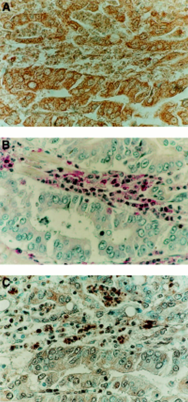
Figure 2 .

Apoptotic lymphocytes adjacent to FasL expressing carcinoma. (A) FasL expression (brown) detected in an area of gastric carcinoma by immunoperoxidase staining. (B) CD45 (leucocyte common antigen) immunophosphatase (alkaline phosphatase conjugated anti-alkaline phosphatase (APAAP)) staining (red) was performed on a sequential section. CD45 positive cells (red) of lymphoid morphology are present adjacent to areas of carcinoma. Slides were counterstained with haematoxylin. Isotype matched control sections were negative (not shown). (C) Cell death detection in situ by terminal deoxynucleotidyl transferase mediated dUTP nick end labelling (TUNEL). Tumour sections sequential to those shown in (A) and (B) were used to detect cell death by enzymic labelling of DNA strand breaks using TUNEL. Only those cells with positive TUNEL staining (brown) and exhibiting apoptotic morphology were considered apoptotic. Control sections (not shown) where the labelling enzyme was omitted were negative.
