
Figure 5 .
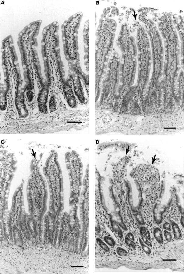
Histological sections from (A) controls, (B) cisplatin (10 mg/kg) treated animals and (C) animals treated with cisplatin 10 mg/kg and ondansetron 300 µg/kg showing degeneration of the tips of the villi and enterocyte damage (arrows). These sections were selected to be representative of the histological appearance for each of the groups. An example of more serious damage after treatment with cisplatin 10 mg/kg is shown in (D), with denudation of the distal third of some of the villi which occurred in 23% of villi in one out of nine animals and was not present in all sections. All sections were examined in a blinded fashion but were `selected' to be representative of the histology in each of the treatment groups. Bar = 100 µm.