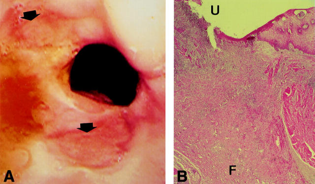
Figure 1 .
(A) Endoscopic image showing two large ulcerations of the distal oesophagus, above the oesophogastric anastomosis (arrows). (B) Microscopic view of surgical specimen (original magnification × 5; haematoxylin and eosin); the ulcer (U) extends to the adventitia with noticeable fibrosis (F).

