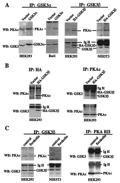
Figure 3.
Physical association between PKA and GSK-3. (A) HEK293, Rat1, and NIH 3T3 were transfected with an empty vector or GSK-3 vectors that express rat GSK-3α (pMT2-GSK3α) or HA epitope-tagged human GSK-3β (pcDNA3- HA-GSK3β). Two days after transfection, cells were starved in serum-free medium for 12 h before lysing. GSK-3 was immunoprecipitated (IP) by using isoform-selective GSK-3α or -β antibodies as described in Materials and Methods. The immunocomplex was analyzed by Western blotting (WB) with an anti-PKAc antibody. The membranes were reprobed with an anti-GSK-3 antibody (reactive with both isoforms) to confirm the efficiency and specificity of immunoprecipitation. The locations of PKAc, GSK-3α, GSK-3β, HA-GSK-3β, and the Ig heavy chain (IgH) are indicated. (B) Lysates from vector or HA-GSK-3β-transfected HEK293 cells were immunoprecipitated with anti-HA monoclonal antibody. The immunocomplex was subjected to Western blotting with an anti-PKAc antibody and then with an anti-GSK-3 antibody as in A. In the reciprocal experiment (Right), PKAc was immunoprecipitated from the same lysates. The immunocomplex was analyzed by Western blotting for GSK-3 and then for PKAc. (C) Untransfected HEK293 and NIH 3T3 cells were starved in serum-free medium for 12–24 h and stimulated with forskolin or vehicle (control) as described in Fig. 1. GSK-3β or PKA RII was immunoprecipitated and the immunocomplex subjected to Western blotting with the indicated antibodies.

