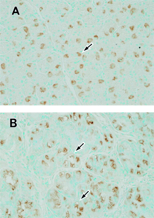
Figure 1 .

Immunohistochemical detection of H+,K+-ATPase in human gastric mucosa. (A) Gastric mucosal tissue of an H pylori negative patient without enlarged folds. Parietal cell cytoplasm is specifically and uniformly stained (arrow). Original magnification × 210. (B) Gastric mucosal tissue of an H pylori positive patient with enlarged fold gastritis. Some parietal cells are uniformly stained as above. In other parietal cells, stained cytoplasm is less uniform and appears granular or contains several vacuole-like clear areas which are negatively stained for H+,K+-ATPase (arrows). Original magnification × 210.
