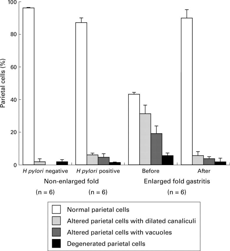
Figure 5 .

Semiquantitative analysis of parietal cell morphology before and after eradication of H pylori in six patients with enlarged fold gastritis and 12 patients without enlarged folds (six H pylori positive and six H pylori negative). Electron micrographs of gastric mucosa were scored according to parietal cell morphology as being normal, altered with dilated canaliculi, altered with vacuoles, or degenerated. The mean of the percentage of each class is represented by bars. Standard errors are represented by vertical lines.
