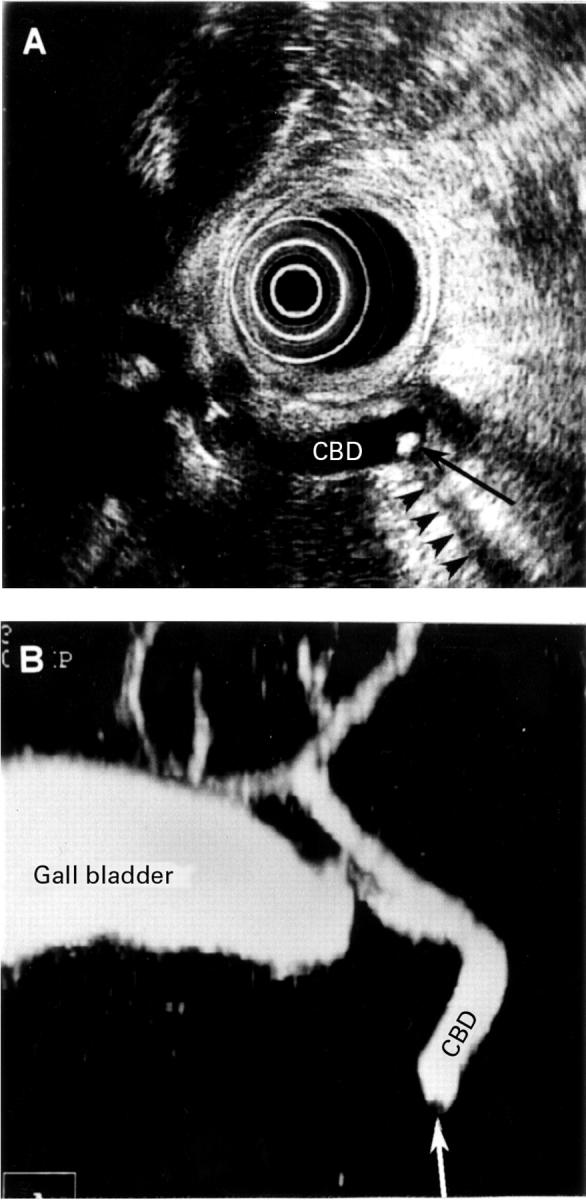
Figure 1 .

Small stone, 4 mm in diameter, in the distal part of undilated common bile duct (CBD). (A) Endosonographic image showing the stone (black arrow) with acoustic shadowing (arrowheads). (B) Helical computed tomographic cholangiography; maximum intensity projection image shows the gall bladder and both the intrahepatic and extrahepatic bile ducts. The small filling defect (white arrow) close to the end of the common bile duct represents the stone.
