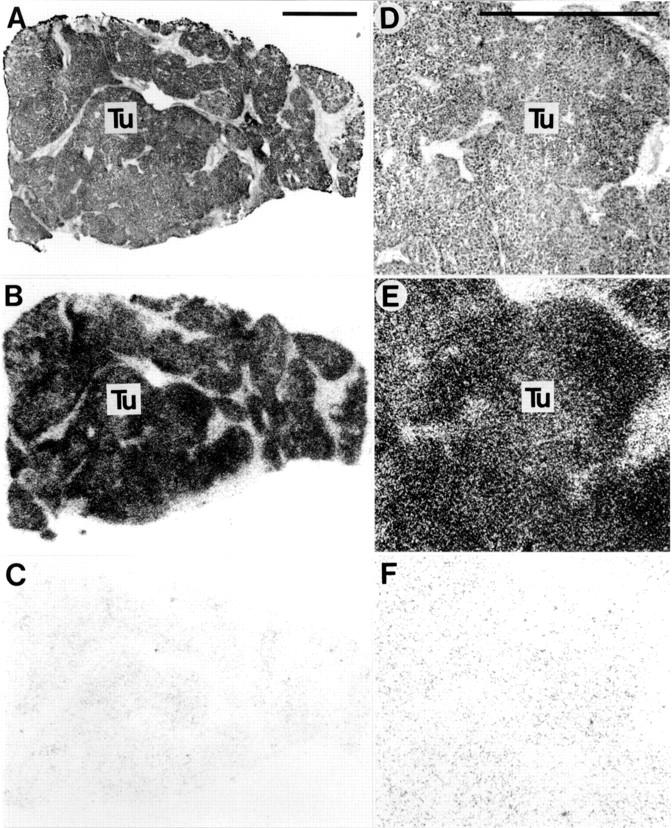
Figure 1 .

Somatostatin receptors in hepatocellular carcinoma. (A,D) Haematoxylin and eosin stained sections. D is an area of A at higher magnification. (B,E) Autoradiograms showing total binding of 125I-labelled [Tyr3]-octreotide. Homogenous labelling of tumour (Tu) tissue can be seen clearly. (C,F) Autoradiograms showing non-specific binding of 125I-labelled [Tyr3]-octreotide (in the presence of 10−6 M unlabelled octreotide). Bars = 1 mm.
