
Figure 3 .
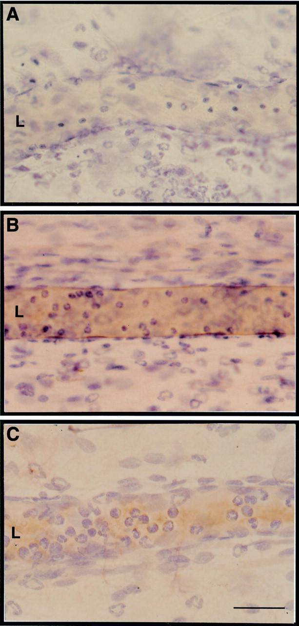
Localisation of intercellular adhesion molecule (ICAM) 1 immunostaining in inflamed rat mesenteric postcapillary venules. Typical micrographs of the ileal section of rat mesentery whole mounts. (A) Preparations excised two hours post-administration of 20 ng rat interleukin (IL) 1β from an animal injected with control mouse IgG 10 minutes prior to sacrifice. (B) As for A, but the rat was injected with 2 mg/kg mouse antirat ICAM-1 10 minutes prior to sacrifice and tissue collection. Note the brown staining around the endothelium of the postcapillary venule. (C) As in B, but the rat was treated with 100 µg/kg subcutaneous dexamethasone one hour prior to intraperitoneal injection of IL-1β. Note the scarce brown immunostaining compared with B. Pictures are representative of five distinct preparations. L, vessel lumen. Bar, 30 µm.